These last 2 months have been a bit of a blur.
1. We were in Nairobi, Kenya, for a 3-week missions training course. It was great to meet other families serving throughout the world and to learn about cross-cultural service.


 But my mind was often taken back to worrying about the well-being of the Eye Hospital in Lusaka which I had left behind. Most of our time in Nairobi was spent in the classroom, but we did have 1 and a half free weekends during which we able to venture out and explore the country.

Nonetheless, we enjoyed our time seeing various animals and birds – and even got to see a lioness with her cubs lounging under a tree. We especially enjoyed watching the antics of the baboons.


The first weekend, we took a day tour of Lake Nakuru. We were expecting to see hundreds, if not thousands of flamingos. But once we got there, we were told by our tour guides that the flamingoes had migrated south to Tanzania for the season and would return in a few months time. Instead, we'd see lots of pelicans. Perhaps they should have told us that before we signed up.

Nonetheless, we enjoyed our time seeing various animals and birds – and even got to see a lioness with her cubs lounging under a tree. We especially enjoyed watching the antics of the baboons.

The second free weekend, we piled 20 people into 4 minivans and caravanned on a 5 hour ride to Masai Mara National Park.

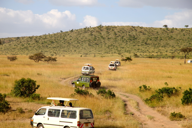

We stayed at the beautiful Sentrim Mara Lodge. We experienced 4 game drives, and each one was super special. So many different animals were spotted, including zebras, many varieties of antelope, buffalo, giraffes, elephants, hippos, warthogs, lions, jackals, hyenas, and even a hunting cheetah! God’s creation is truly magnificent.


One of the highlights of the trip was getting to sleep in the hotel “tents” at the lodge. Throughout the night, we heard the howling of hyenas and groans of cheetahs; it was truly an incredible experience sleeping amidst the wild animals.
For more animal photos, feel free to see the Facebook Kenya album.
For more animal photos, feel free to see the Facebook Kenya album.
2. We fired our maid. Although it was sad to lose a good helper, we realized it was just too stressful for us to leave someone alone in our home all day, especially if we were not able to fully trust her. Hopefully we can find another helper soon.
3. We’ve been working with a wonderful Canadian donor, Colin Glassco, to sponsor one day of children’s surgeries every month. This has been a welcome relationship, in which the neediest patients who cannot afford to pay for surgery have been able to undergo surgeries free of charge on the specific Glassco days.


 4. I had another opportunity to attend another National Prevention of Blindness Committee meeting, specifically addressing the issue of trachoma in Zambia. It was great to sit with other ophthalmologists, representatives from the International Trachoma Initiative, and country directors of donor organizations like Sightsavers International, CBM, and Operation Eyesight Universal, to discuss the problem of trachoma in Zambia and figure out ways to tackle the issue.


5. Last week, Zambia made history! Zambia won the African cup of nations (soccer) for the first time in a close match against Ivory Coast! The whole country was in a state of celebration.
6. I was on local television twice! Although I never saw the news report, nor do I receive “Muvi TV” on my television set, I have heard that the interviews were aired. Unlike American TV shows, which are taped weeks ahead of time, edited, and then finally aired, Muvi TV and other local African channels tape and air shows on the same day. The whole situation came about because a small baby with sclerocornea had parents who were very proactive in finding a sponsor for their child’s corneal transplant. They sought out a TV channel and were interviewed by Muvi TV. On television, they explained their plight – they did not have the finances to fund a corneal transplant. A local Lions Club approached me and asked if they would partner with me in helping this baby undergo surgery. We agreed and set a date for the surgery. Muvi TV interviewed me and the patient’s family preoperatively, and that was aired a month ago. Last week, Muvi TV came to film the actual surgery and another interview. Apparently, this, too, was aired last week.
WARNING! Please stop reading now if you are averse to seeing my eye photos.
7. Another batch of corneas arrived!
 Midwest Eye Banks was able to send another set of fresh corneal tissues, so I did 5 transplants last week.


Patient #1 was the sclerocornea baby. She was 2 years old and responsive only to light in both eyes since birth.
I performed the corneal transplant in the right eye, but not with a struggle. As soon as I opened the eye, I realized the iris and lens were stuck to the cornea. I had no choice but to remove everything and to leave the child aphakic. The process of sewing a graft onto the squishy 2-year-old’s sclera was akin to sewing together two soft, rotting grapes. But somehow I managed to close the globe.


And somehow, the next day, the child was able to track my finger with her new eye! Her parents were delighted that she seemed to be "seeing" for the first time. She was very sensitive to light, so most of the photos I snapped of her captured the back of her head. Here's one blurry shot of her face.

Patient #2 was a 43-year-old man with Mooren’s ulcers. His left eye was essentially hopeless, with iris bulging out and no real corneal substance left to support the globe. We had scheduled him for an evisceration. However, his right eye seemed to have some slight hope. He had pockets of iris pooching out in 3 places inferiorly. There was only a small area of firm cornea intact superiorly.


I realized the only way to help him was to perform a large-diameter graft. Although it was difficult to find and clean up a firm area on the inferior sclera onto which to anchor the donor corneal tissue, after much intraoperative prayer I managed to close the eye. And although his inferior iris was chopped off, fortunately his lens remained intact.


He was my happiest patient on postoperative day #1. He was able to see CF@2 meters and was finally able to walk around on his own without being led by the hand.


Patient #3 was a 28-year-old girl who was phthisical in her right eye and had a dense corneal scar in her left (HM vision). I was not too clear about her visual prognosis, but we agreed to attempt surgery.
 Fortunately, her lens was not cataractous, so I left it intact. Also, the iris was only adherent to the cornea inferiorly, so I was able to leave the superior part intact.

Postop day #1, she was able to CF@1.5 meters and slowly walking around on her own. She seemed pleased with that.
 Patient #4 was the mother of one of our hospital employees. She had been blind in both eyes for quite some time, and both corneas were very swollen. Again, knowing the guarded visual prognosis, I did not guarantee any promise of success.

Intraoperatively, her iris and lens were in no state to keep, so I was forced to cut them out with scissors. What was left of the lens was really a fibrosed lens capsule, as there was no evidence of a crystalline lens anywhere to be found. My heart sank when I realized that leaving her aphakic was my only option. Nonetheless, I sutured up the eye and prayed for the best.

On postop day #1, her cornea was very swollen, and her vision was no better (HM preop, HM postop). Hopefully, she’ll get some improvement once the corneal edema and vitreous heme subside. Only time will tell.


Postop day #1, she was able to CF@1.5 meters and slowly walking around on her own. She seemed pleased with that.


Intraoperatively, her iris and lens were in no state to keep, so I was forced to cut them out with scissors. What was left of the lens was really a fibrosed lens capsule, as there was no evidence of a crystalline lens anywhere to be found. My heart sank when I realized that leaving her aphakic was my only option. Nonetheless, I sutured up the eye and prayed for the best.

On postop day #1, her cornea was very swollen, and her vision was no better (HM preop, HM postop). Hopefully, she’ll get some improvement once the corneal edema and vitreous heme subside. Only time will tell.
Patient #5 was supposed to be my slam-dunk patient. She was a 20-year-old female with keratoconus of both eyes – large Vogt striae across the middle of her corneas and central corneal scarring.

She wore hard contact lenses, and her best vision with those lenses was 6/36 in both eyes. Without glasses or contacts, she was CF@1 meter. She was quite myopic with some astigmatism, so I decided to oversize the donor cornea only 0.25 mm larger than the host bed (instead of the usual 0.5 mm) to give her some corneal flattening. That was my first mistake. My second mistake was to re-use an autoclaved trephine to cut the donor tissue. An incomplete cut required a manual completion using corneal scissors, resulting in a slightly irregular edge for ¼ of the donor tissue perimeter. These factors combined led to a 2 hour surgery in which I struggled to close the eye. It just kept leaking and leaking, and I kept adding suture after suture. Instead of my normal 16 suture transplant, I ended up placing 27 in this poor patient. Finally, after what seemed like eternity, I managed to close the eye enough to hold a chamber.

She wore hard contact lenses, and her best vision with those lenses was 6/36 in both eyes. Without glasses or contacts, she was CF@1 meter. She was quite myopic with some astigmatism, so I decided to oversize the donor cornea only 0.25 mm larger than the host bed (instead of the usual 0.5 mm) to give her some corneal flattening. That was my first mistake. My second mistake was to re-use an autoclaved trephine to cut the donor tissue. An incomplete cut required a manual completion using corneal scissors, resulting in a slightly irregular edge for ¼ of the donor tissue perimeter. These factors combined led to a 2 hour surgery in which I struggled to close the eye. It just kept leaking and leaking, and I kept adding suture after suture. Instead of my normal 16 suture transplant, I ended up placing 27 in this poor patient. Finally, after what seemed like eternity, I managed to close the eye enough to hold a chamber.
But somehow, miraculously, on postop day #1, despite her significant corneal edema, she was seeing 6/36!

There are so many times that the Lord has made up for my deficiencies due to my complications in less than ideal surgical situations with less than ideal equipment/microinstruments. I praise God that despite my inadequacies, He covers so many of my mistakes and helps my feeble efforts as I do my best to help my patients.

There are so many times that the Lord has made up for my deficiencies due to my complications in less than ideal surgical situations with less than ideal equipment/microinstruments. I praise God that despite my inadequacies, He covers so many of my mistakes and helps my feeble efforts as I do my best to help my patients.